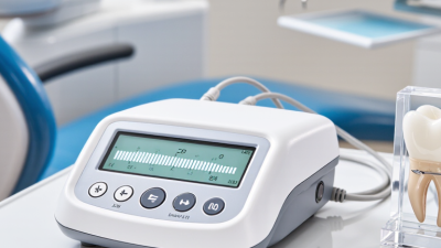
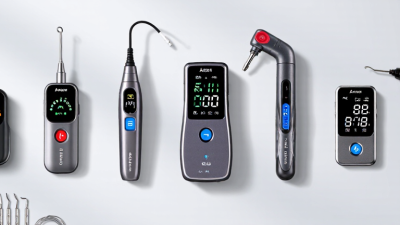
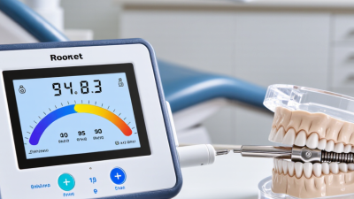
Choosing an apex locator is not a box-checking exercise. With the apex locator woodpex v, dentists should assess how clearly readings appear, how easily the unit fits into a treatment setup, and whether its instructions match the team’s workflow. A bright display matters under operatory lighting. So does a stable clip connection when the patient moves or the canal is damp. Small details can shape daily use.
This overview will examine the Woodpex V through practical questions: What does the device show during canal measurement? How straightforward is setup? Which limitations should clinicians consider before relying on its readings? Electronic measurements support working-length decisions, but they do not replace clinical judgment or other appropriate checks. Canal anatomy, irrigants, and contact conditions may affect readings. Real cases are rarely as tidy as product photos suggest.
A useful reminder: “A reading informs; judgment confirms.” I cannot verify this wording as a quotation from a named dental expert, so I will not attribute it to one. A fabricated expert quote would weaken trust rather than strengthen it. Dentists comparing the apex locator woodpex v should consult the current manufacturer instructions, assess the device in their own clinical setting, and consider feedback from qualified colleagues. This article aims to make that evaluation more concrete, while recognizing that no single device suits every practice.
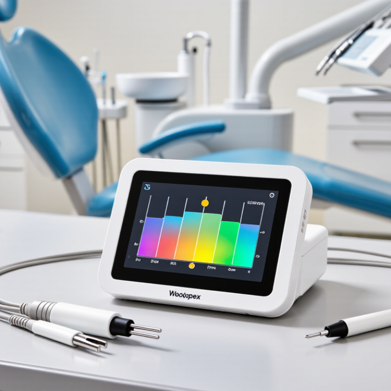
An electronic apex locator estimates canal length by measuring electrical impedance between a file inside the canal and an electrode attached to the patient. As the file approaches the apical constriction, the relationship between impedance readings at different frequencies changes. The device converts that change into a moving display or audible signal. It does not directly “see” the root tip. A clinician still needs to interpret the reading alongside anatomy and clinical findings.
A 2004 review by Gordon and Chandler in the International Endodontic Journal summarized studies reporting roughly 90% accuracy within ±0.5 mm, while noting that results varied with canal conditions. That tolerance matters: the anatomical apex and apical constriction are not always in the same place. Saliva, irrigant, a wide open apex, or file contact with metal can also affect readings. Keep the chamber reasonably dry, use a stable file, and recheck an unexpected measurement. Small details count. An apex locator is useful, but not infallible; even an experienced operator may need to pause when readings fluctuate.
An electronic apex locator can help dentists estimate the canal’s endpoint, but the AAE’s 0.5–1.0 mm apical reference is a clinical guide, not a universal measurement. Working length is commonly set short of the anatomical apex to help keep cleaning and filling within the canal. The distance between the radiographic apex, apical foramen, and constriction can vary. That matters. A locator reading should be interpreted alongside the tooth’s anatomy, canal conditions, and a confirming radiograph when indicated.
A stable reading is more useful than a rushed one. Keep the canal appropriately moist, avoid flooding the chamber, and check that the file can move freely. If readings fluctuate, pause and inspect the contact, file position, and canal contents. Recheck after adjustments. An electronic signal does not reveal every anatomical detail, especially in irregular or resorbed roots. Clinical judgment still matters, and occasionally the first measurement needs revision.
Tips: Advance the file slowly and watch for a consistent display near the apical reference. Record the chosen working length and reassess it if the file, reference point, or canal conditions change. Small differences can be meaningful. Don’t treat a single beep as proof; compare the finding with the rest of the clinical picture.
| Assessment dimension | Clinical reference | How to use it | Important limitation |
|---|---|---|---|
| Working-length reference | A commonly used target is approximately 0.5–1.0 mm short of the apical foramen. | Use this range as a clinical reference when interpreting electronic measurements and confirming the intended treatment length. | It is a general reference, not a fixed endpoint for every tooth or canal. |
| Apical constriction | The narrowest part of the apical canal, when present, is often near but not necessarily identical to the apical foramen. | Interpret the electronic reading alongside canal anatomy, tactile feedback, and imaging rather than treating a display marker as a direct view of the constriction. | The constriction may be absent, irregular, or difficult to identify, especially in immature or resorbed roots. |
| Electronic measurement | Electronic apex locators estimate file position in relation to the canal terminus using electrical measurements. | Advance the file gradually and look for a stable, repeatable reading; record the measurement and reassess if readings fluctuate. | An electronic reading is an estimate and should be interpreted with the clinical findings and appropriate radiographic information. |
| Radiographic assessment | A periapical radiograph can show the file’s projected position relative to the root outline and radiographic apex. | Use imaging when clinically indicated to assess anatomy, confirm file position, or investigate disagreement between findings. | A two-dimensional image may not show the foramen’s exact location; angulation and root curvature can affect interpretation. |
| Canal conditions | Excess fluid, conductive materials, or an unstable file connection can interfere with consistent measurements. | Follow the device instructions, maintain a suitable canal condition, and check the file clip, lip clip, and electrical connections. | Do not assume that every inconsistent reading indicates an anatomic problem; first check setup and operating conditions. |
| Repeatability check | Repeated measurements should be reasonably consistent under the same clinical conditions. | Withdraw and re-advance the file as appropriate, then compare readings before finalizing the working length. | Repeatability supports confidence but does not by itself prove the exact position of the apical constriction. |
| Clinical decision | Working length is determined from the combined assessment of anatomy, electronic measurement, imaging when appropriate, and clinical judgment. | Document the selected length and the evidence used to establish it; reassess if anatomy or treatment findings change. | The appropriate endpoint is case-specific and should be determined by a qualified dental professional. |
Clinical note: The 0.5–1.0 mm reference is a guide for working-length assessment, not a guarantee of anatomical accuracy or a substitute for case-specific clinical judgment.
Contemporary endodontic workflows need dependable measurements, clear feedback, and steady infection control. An electronic apex locator provides real-time visual and audible cues as a file advances through the canal. This can reduce repeated length checks and help clinicians coordinate measurement with radiographic review. WHO’s Global Oral Health Status Report (2022) estimates that untreated caries in permanent teeth affects about 2 billion people worldwide. That burden makes efficient, tooth-preserving care important, though it does not prove that any single device improves outcomes.
Useful features are those that fit the appointment, not merely those that look advanced. A readable display, stable signal, and simple controls can support work under magnification while the clinician maintains isolation. Record the measurement and reassess when anatomy, symptoms, or readings seem inconsistent. Irrigants, canal shape, and file contact can affect signals. A number on a screen is not a diagnosis, and radiographs still have a role.
Tips: Keep the canal conditions consistent, use a compatible file, and pause if the reading jumps. Verify unexpected measurements before shaping. Small interruptions matter.
An apex locator should be evaluated against the ±0.5 mm accuracy benchmark using a defined reference point, not display appearance alone. The key question is whether its readings consistently place the file tip within half a millimeter of the established apical endpoint. A clear screen helps, but it cannot prove accuracy. Small differences matter.
Testing should include repeated measurements in extracted teeth or suitable laboratory models, with the reference length verified independently. Record how often readings fall within the benchmark, how widely results vary, and whether measurements remain stable after repositioning the file. Include different canal conditions, since moisture, irrigants, and contact with metallic restorations may affect readings. Keep the procedure consistent. A single successful reading is weak evidence.
In daily practice, compare electronic readings with other clinical findings and follow the device’s instructions. Check that the file clip and lip electrode are secure, and investigate erratic signals rather than accepting them automatically. Not a perfect test. Laboratory results may not predict every patient’s anatomy, and operator technique can introduce variation. A useful evaluation reports both accuracy and repeatability, while stating the testing conditions and limitations plainly.
The bar shows the stated ±0.5 mm deviation benchmark around the target endpoint. It represents an evaluation criterion, not comparative clinical test results for any device.
Radiographic verification is most useful when the canal anatomy or measurement conditions make an electronic reading less dependable. Examples include open apices, resorption, perforation, unusual curvature, or a reading that changes repeatedly as the file advances. A stable display is helpful, but it does not show the full root shape. Take a working-length radiograph when the electronic measurement conflicts with tactile findings or the expected anatomy.
Evidence supports using both methods as complementary checks, not treating either as infallible. A clinical systematic review by Martins and colleagues in the International Endodontic Journal found that electronic apex locators can determine working length reliably, while also noting variation among studies and clinical conditions. The European Society of Endodontology’s 2023 S3 guideline likewise frames treatment decisions around the available clinical and radiographic evidence. A small but important detail: the file tip must be visible and positioned clearly against the root image.
For a routine case, record the electronic reading, confirm file position radiographically when indicated, and reassess if the two disagree. Check the image for distortion, overlap, or an unclear apex before changing the working length. Don’t force agreement. A radiograph is a two-dimensional view, and an electronic reading can also be affected by fluid, contact, or canal anatomy. When the evidence remains uncertain, pause and review the anatomy rather than relying on a single number.
DENTSPLY Maillefer - USA
5100 E. Skelly Drive, Suite 300
Tulsa, Oklahoma 74135
DENTSPLY Maillefer - Canada
161 Vinyl Court
Woodbridge, ON L4L 4A3
Toll-free in the U.S.:
1-800-924-7393
Toll-free in Canada:
1-800-263-1437
Toll-free in the U.S.:
1-800-924-7389
Toll-free in Canada:
1-888-336-8775