What Is Cell RNA Extraction and How Does It Work?
Cell Rna Extraction is the laboratory process of isolating RNA from cells for careful molecular analysis. It helps researchers examine gene activity, cellular responses, and disease-related changes. Unlike DNA, RNA is fragile and breaks down quickly when exposed to heat, moisture, or RNase enzymes. Small details matter.
In a typical workflow, researchers collect cells and disrupt their membranes with a specialized lysis solution. This releases RNA while helping deactivate destructive enzymes. The mixture then undergoes separation, often through a silica column or magnetic beads. RNA binds to the selected surface, while proteins, lipids, and other cell materials are removed during washing. A final elution step releases purified RNA into a small tube. The liquid may look clear, but appearance proves little.
Reliable extraction requires RNase-free equipment, clean handling, and validated procedures. Researchers commonly measure RNA concentration and purity with spectrophotometry or fluorescence-based tools. They may also examine RNA integrity before sequencing or reverse transcription. Appropriate controls can reveal contamination, degradation, or unexpected sample loss. The process is not flawless. A high concentration reading can still hide damaged RNA. That assumption can fail.
Experienced laboratory teams compare results with quality thresholds, documented protocols, and instrument controls. They also follow the instructions supplied with extraction kits and maintain consistent sample temperatures. When performed carefully, Cell Rna Extraction produces RNA suitable for downstream research. Its success depends less on one dramatic step than on disciplined handling throughout the workflow.
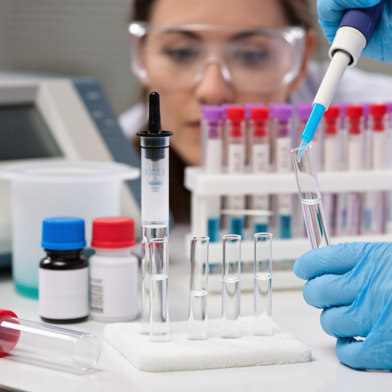
Cell Harvesting and Lysis: Releasing RNA While Limiting Cellular RNases
What Is Cell RNA Extraction and How Does It Work?
Cell RNA extraction begins with careful harvesting. Keep cells cold and process them quickly. Warm delays can activate cellular RNases, enzymes that break RNA into smaller fragments. Remove culture fluid, wash gently, and collect the cells without creating unnecessary foam. A clean pellet helps reduce salts, proteins, and leftover medium that may interfere with later analysis.
Cell lysis releases RNA by breaking the cell membrane and weakening protein structures. A suitable lysis solution often contains compounds that disrupt membranes and inactivate RNases. Mix thoroughly, but avoid harsh handling that can shear RNA or create uneven results. For dense pellets, brief homogenization may improve recovery. In practice, this step is easy to underestimate. Even careful work can leave a small compromise between complete lysis and RNA damage.
Tips: Use RNase-free tubes, tips, and work surfaces. Change gloves after touching shared equipment. Keep samples cold before lysis, and avoid repeated freeze-thaw cycles. Check the lysate visually for clumps. If material remains, adjust mixing gently rather than forcing it. Cell type, sample size, and lysis chemistry all affect performance, so a small pilot test is often worthwhile. Mistakes happen; documenting timing and handling conditions makes the next extraction more reliable.
What Is Cell RNA Extraction and How Does It Work?
Approximate RNA composition in a typical mammalian cell
During cell harvesting and lysis, membranes are disrupted to release cellular RNA. Rapid processing, cold conditions, and RNase-inhibiting lysis reagents help protect the RNA from degradation. Ribosomal RNA generally represents the largest fraction of total cellular RNA, while messenger RNA is a much smaller fraction.
Values are approximate averages for mammalian cells and can vary with cell type, growth state, sample handling, and extraction method.
RNA Purification: Silica Membranes and 70–80% Ethanol Washing
Cell RNA extraction depends on a simple chemical shift: RNA binds silica when chaotropic salts remove its hydration shell. In a small spin column, the lysate passes through a porous membrane. RNA stays behind. Proteins, lipids, and cell debris move away.
The ethanol wash is more important than it looks. A 70–80% ethanol solution removes salts while preserving RNA attachment to silica. Too little ethanol can release RNA prematurely. Too much may leave residual alcohol after centrifugation. That residue can inhibit reverse transcription and distort quantitative PCR results. I usually add a second dry spin. It feels excessive, but wet membranes cause many avoidable failures.
Keep the membrane clean. Avoid disturbing the cloudy lysate pellet. After washing, let the column air-dry briefly, then elute with low-salt, nuclease-free water. Elution works best when the liquid contacts the membrane evenly. The 2006 BMC Molecular Biology study introduced the RNA Integrity Number, or RIN, using a 1–10 scale for RNA quality assessment. The MIQE guidelines in Clinical Chemistry also stress recording RNA quantity, purity, and integrity before amplification. A high concentration is not proof of good RNA. I have seen clear eluates perform poorly because ethanol remained. Spectral readings alone can be misleading.
DNase Treatment: Removing Genomic DNA Without Degrading RNA
What Is Cell RNA Extraction and How Does It Work?
Cell RNA extraction separates fragile RNA from proteins, lipids, and genomic DNA. DNase treatment is often the quiet quality-control step. DNase I digests DNA by cutting its phosphodiester backbone, while RNA remains protected under suitable conditions. It does not remove RNA itself.
Genomic DNA can inflate RNA concentration readings and create false signals in reverse-transcription PCR. The MIQE guidelines, published in Clinical Chemistry, identify DNA contamination controls as essential for reliable quantitative PCR. The RIN integrity scale ranges from 1 to 10, but a high score cannot prove that DNA is absent. I always check both.
DNase may be applied during extraction or after purification. On-column treatment is convenient, while in-solution treatment can improve contact with residual DNA. Enzyme exposure, buffer pH, temperature, and incubation time must match the protocol. Excessive treatment is not automatically safer. It can increase handling loss and leave cleanup problems. My own practice is slightly cautious: I include a no-reverse-transcription control, even when the sample looks clean.
Tips: Use nuclease-free tubes and low-retention tips. Mix gently, not violently. After treatment, remove the enzyme and salts completely. Measure RNA with a fluorometric method when possible, then inspect purity ratios. A no-RT control should remain negative. If it amplifies, DNase treatment was incomplete—or the assay needs reconsideration. That small failure can reveal more than a perfect-looking concentration value.
Sources: MIQE Guidelines, Clinical Chemistry, 2009; RNA integrity standards reported in analytical electrophoresis literature.
RNA Elution and Downstream Use: Supporting RT-qPCR, RNA-Seq, and Microarrays
After cell lysis and purification, RNA elution releases the captured molecules into a small volume of RNase-free solution. The goal is simple: recover clean RNA without damaging its integrity. A warm elution buffer can improve recovery, but excessive heat may increase degradation. Gentle pipetting helps. Vigorous mixing can shear fragile RNA.
Eluted RNA supports several downstream applications. For RT-qPCR, concentration and purity affect reverse transcription and amplification consistency. Residual genomic DNA can create misleading signals, so DNase treatment may be necessary. RNA-seq requires stronger quality control because fragmented RNA can distort transcript coverage. Microarrays also depend on intact, representative RNA for reliable hybridization. Measure concentration with a suitable method, then check purity ratios and integrity when possible. A clear eluate is not proof of quality. I have learned that small handling delays can matter more than expected.
Tips: Keep samples cold and use low-retention, RNase-free tubes. Elute with the smallest practical volume, but avoid concentrating contaminants. Include a no-template or no-reverse-transcription control when appropriate. Record elution volume, concentration, and storage time. If RNA quality looks acceptable but results remain inconsistent, reassess cell input, lysis timing, and freeze-thaw exposure.
